Study Shows Activation of Pulmonary Macrophages by SARS-CoV-2 Spike Glycoprotein
02/23/2023
In a recent study published in the International Journal of Molecular Sciences, researchers investigated the effects of severe acute respiratory syndrome coronavirus 2 (SARS-CoV-2) spike (S) glycoprotein on human lung macrophage (HLM) activation and angiotensin-converting enzyme 2 (ACE2) and transmembrane serine protease 2 (TMPRSS2) expression in human lung macrophages.
The coronavirus disease 2019 (COVID-19) is characterized by airway inflammation. The etiological agent of COVID-19, SARS-CoV-2, enters host cells by binding to the ACE2 receptors, utilizing TMPRSS2. Macrophages are most abundantly present immunological cells in pulmonary tissues and are involved in several functions regulated by chemokine and cytokine production.
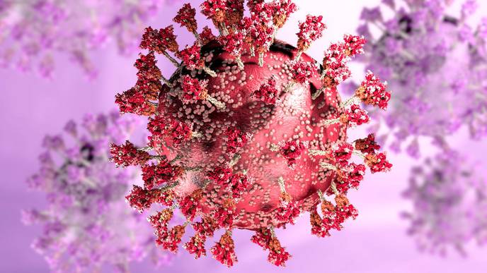
Study: SARS-CoV-2 Spike Protein Activates Human Lung Macrophages. Image Credit: piccreative / Shutterstock
About the study
In the present study, researchers evaluated SARS-CoV-2 S glycoprotein’s effects on inflammatory chemokine and cytokine release by human lung macrophages and TMPRSS2 and ACE2 expression in the macrophages.
The human lung macrophages were purified from pulmonary tissues of individuals with hepatitis C virus (HCV)-positive, hepatitis B virus (HBV) surface antigen-negative, and human immunodeficiency virus (HIV)-negative individuals undergoing pulmonary tissue resection. The effects of increasing concentrations of SARS-CoV-2 S on the activation of HLMs were evaluated in cell culture experiments, wherein human lung macrophages were stimulated with lipopolysaccharide (1.0 µg per mL), or increasing SARS-CoV-2 S (0.010 µg per mL to 10.0 µg per mL).
MTT [3-(4,5-dimethylthiazol-2-yl)2,5-diphenyl tetrazolium bromide] assays were performed to assess viable HLM proportions. The levels of mediators [interleukin (IL)-1β, IL-6, tumor necrosis factor-alpha (TNF-α), CXCL-8, angiopoietin 1 (ANGPT1), vascular endothelial growth factor A (VEGF-A), angiopoietin 2, and tumor growth factor-beta (TGF-β)] in the culture supernatants were evaluated using enzyme-linked immunosorbent assays (ELISA).
TMPRSS2, ACE2, IL-1β, C-X-C motif ligand 8 (CXCL-8), TNF-α, and IL-6 messenger ribonucleic acid (mRNA) levels were measured using quantitative reverse-transcription-polymerase chain reaction (RT-qPCR). The intracellular cytosolic and lysosomal intracellular calcium ion levels were determined using computer-assisted video imaging at the single-cell level.
Phagocytosis assays were performed using E. coli strains, and high-content and time-lapse microscopic analyses were performed to investigate whether SARS-CoV-2 S can induce phagocytosis by HLMs. In addition, HLM morphology and tracking were assessed. To investigate whether toll-like receptor 2 (TLR-2) expression contributed to SARS-CoV-2 S-induced human lung macrophage activation, HLMs were preincubated with antibodies against TLR-2 followed by S glycoprotein stimulation.
Results
SARS-CoV-2 S induced the secretion of inflammatory chemokines and cytokines such as TNF-α, IL-1β, IL-6, and CXCL-8 release from primary human lung macrophages, with significant effects at 1.0 µg per mL and 10.0 µg per mL, not regulated by toll-like receptor 2. The S glycoprotein promoted phagocytosis and increased intracellular calcium ion concentration in the lysosomes of human lung macrophages and affected human lung macrophage tracking by inducing cellular displacement changes; however, HLM morphology remained unaffected.
Human lung macrophages constitutively expressed messenger ribonucleic acid for angiotensin-converting enzyme 2 and transmembrane serine protease 2 (to a greater extent), indicating that the human lung macrophages are S glycoprotein targets. However, the proportion of viable human lung macrophages after 18 hours of S glycoprotein treatment was no different from that within the untreated lung macrophages.
The findings indicated that the S glycoprotein induced the release of chemokines and cytokines from human lung macrophages but not their de novo synthesis and that the toll-like receptor 2 was not needed for the release. Concerning human lung macrophage tracking, S glycoprotein-stimulated macrophages were situated nearer the origination point and assimilated in smaller displacement areas than untreated cells; however, HLM speed remained unaffected.
SARS-CoV-2 S glycoprotein-activated human lung macrophages showed greater uptake of Escherichia coli particles than controls. The S glycoprotein regulated the intracellular calcium ion concentrations without any cytotoxic effects on human lung macrophages. SARS-CoV-2 S glycoprotein-stimulated lysosomal calcium ion accumulation could be due to interactions with acidic cellular organelles.
Conclusions
Based on the study findings, human lung macrophages may be considered a double-edged sword during COVID-19. The macrophages may activate phagocytic processes and eliminate pathogenic microbes and cellular debris. However, on the other hand, human lung macrophages may contribute to pulmonary damage, immune dysfunction, and respiratory diseases by enhancing the release of pro-inflammatory substances.
The study findings may elucidate the hyperinflammatory cytokine storm observed in severe SARS-CoV-2 infections. Furthermore, the detection of the ligands and sensors associated with COVID-19-associated cytokine excess may aid in the development of targeted COVID-19 therapeutics to reduce the global health burden of SARS-CoV-2 infections.
Journal reference:
- Palestra, F.; Poto, R.; Ciardi, R.; Opromolla, G.; Secondo, A.; Tedeschi, V.; Ferrara, A.L.; Di Crescenzo, R.M.; Galdiero, M.R.; Cristinziano, L.; et al. SARS-CoV-2 Spike Protein Activates Human Lung Macrophages. Int. J. Mol. Sci. 2023, 24, 3036. DOI: https://doi.org/10.3390/ijms24033036, https://www.mdpi.com/1422-0067/24/3/3036
Facebook Comments